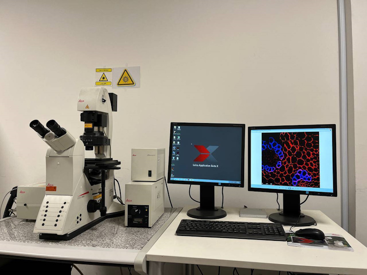
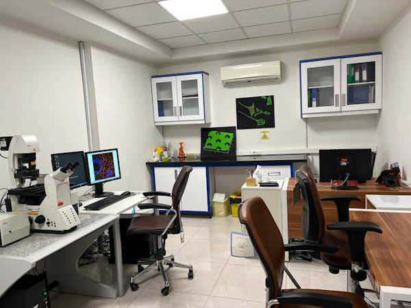
In order to introduce and familiarize more with the laboratories of Sharif University of Technology; Get to know the activities and services of the confocal laser scanning microscopy laboratory.

According to the public relations report of Sharif University of Technology, the Confocal Microscope Laboratory was established in 2017, and a significant amount of students of Sharif University of Technology in the fields of physics, materials, nano, etc. use this laboratory daily, and on average, 100 people annually Students and researchers inside and outside the university benefit from the services of this laboratory. This laboratory is located in the complex of Professor Reza Rusta Azad, located in the laboratory service center of Sharif University of Technology and is always ready to serve students, researchers and industrialists.
Confocal microscope or laser confocal scanning microscope is a microscope that uses the fluorescence properties of the sample to image it, and it has received a lot of attention today.
One of the important applications of confocal microscope is in the imaging of cells and biological samples. The structure of cells and its specific parts can be seen with high magnification and quality after staining with fluorescent material.
Confocal laser scanning microscopy or confocal microscopy (CLSM) is an optical microscopy method that is used for three-dimensional imaging of samples with fluorescent markers.
The key feature of the confocal microscope is its ability to create high-resolution and high-contrast images of thick, transparent samples at different depths.
In this microscope, light sources in the visible region are used to excite the sample, and after refinement, confocal fluorescence images are made using adjustable optical filters.
The ability to penetrate into the depths of the sample with high resolution and contrast and the possibility of layer-by-layer photography (Z-Scan) and the possibility of forming a three-dimensional image of the structure of the sample are among the features of confocal laser scanning microscope.
Confocal analysis CLSM microscope (Confocal Laser Scanning Microscopy) has the ability to perform microscopy in two ways, confocal and fluorescence. In fluorescence microscopy, the entire sample is excited by light passing through a special color filter, and the emitted light forms the desired image, just like in bright field microscopy. In the fluorescence microscopic method, the resolution power is reduced due to the overlap of light emitted from different points.
Confocal microscopy is a solution to compensate for this reduction in resolution; In this type of microscopy, the laser is focused on a specific small area of the sample, and using a special optical system, the emitted light emitted only from that point is recorded by a photodiode, which increases the resolution of the image.
Advantage and application of confocal microscope
This microscopic method has become a valuable tool for research in the field of biological and medical sciences. For example, this method is used for imaging thin optical sections in living and fixed samples with a thickness of up to 100 micrometers, and researchers and industrialists in the mentioned fields use this microscope.
In wide-field fluorescence microscopes, the excited light is irradiated to the entire sample at the same time, and as a result, there is a possibility of exciting the fluorescence molecules throughout the sample. For this reason, data collection in these microscopes is done at a high speed. But exposing the entire sample to light can excite fluorescent molecules that are located in unwanted parts of the sample (usually outside the optical focus), resulting in a large amount of excess background light.
Confocal microscopy offers several advantages over conventional wide-field light microscopy, including:
Ability to control depth of field
Increasing resolution by removing or reducing background information away from the focal plane (factors that degrade image resolution)
Ability to collect serial optical sections (Z Stack) from thick samples (without physical cutting) and form a 3D image
Imaging several different color channels at the same time
For more information and to contact the laboratory office, those interested can call 66166246 (100).

